การตรวจภายใน

มะเร็งเป็นโรคที่คร่าชีวิตคนไทยเป็นอันดับต้นๆ การวินิจฉัยได้เร็วจะทำให้การรักษาได้ผลดี ผู้ชายมักจะเป็นมะเร็งในที่ซ่อนเร้นทำให้เมื่อเกิดอาการโรคก็ลุกลามจนยากจะเยียวยาเช่น มะเร็งปอด มะเร็งตับ แต่สำหรับผู้หญิงนับว่าโชคดีเพราะเป็นมะเร็งในที่สามารถตรวจได้เร็ว เช่น มะเร็งเต้านม มะเร็งปากมดลูกเรื่องมะเร็งเต้านมก็สามารถคลำได้ด้วยตัวเอง ส่วนมะเร็งปากมดลูกก็มีการตรวจ Pap test ซึ่งสามารถให้การวินิจฉัยได้อย่างรวดเร็ว เรามารู้จักกันว่า Pap test เป็นอย่างไร

ใครที่ต้องตรวจ Pap test

แน่นอนว่าต้องเป็นผู้หญิงและต้องอยู่ในวัยเจริญพันธุ์ด้วย สมาคมโรคมะเร็งของอเมริกาได้แนะนำให้เริ่มตรวจ Pap หลังจากมีเพศสัมพันธ์ครั้งแรกไปแล้ว 3 ปี หรือมีอายุมากกว่า 21 ปีแม้ว่าจะไม่มีเพศสัมพันธ์ก็ให้ตรวจตามตารางข้างล่าง

อายุ ความถี่ของการตรวจ
21 - 29 ให้ตรวจปีละครั้ง
30 - 69 ให้ตรวจทุก 2-3ปีหากการตรวจ 3 ครั้งหลังให้ผลปกติ
70 และมากกว่า ให้หยุดตรวจเมื่อการตรวจ 3 ครั้งหลังและ 10 ปีที่ผ่านมาผลการตรวจปกติ

คำแนะนำของอเมริกา

  • All women should begin cervical cancer testing (screening) about 3 years after they start having sex (vaginal intercourse). A woman who waits until she is over 18 to have sex should start screening no later than age 21. A regular Pap test should be done every year. If the newer liquid-based Pap test is used, testing can be done every 2 years.
  • Beginning at age 30, women who have had 3 normal Pap test results in a row may be tested less often - every 2 to 3 years. Either the conventional (regular) Pap test or the liquid-based Pap test can be used.
  • Another reasonable option for women over 30 is to get tested every 3 years (but not more frequently) with either the regular Pap test or liquid-based Pap test, plus the HPV DNA test (see below for more information on this test).
  • Women who have certain risk factors should continue getting tested yearly. This includes women exposed to diethylstilbestrol (DES) before birth and those with a weakened immune system (from HIV infection, organ transplant, chemotherapy, or chronic steroid use).
  • Women who have had a total hysterectomy (removal of the uterus and cervix) may choose to stop having cervical cancer testing, unless the surgery was done as a treatment for cervical cancer or pre-cancer. Women who have had a hysterectomy without removal of the cervix (simple hysterectomy) need to continue cervical cancer screening, and should continue to follow the guidelines above.
  • Women 70 years of age or older who have had 3 or more normal Pap tests in a row and no abnormal Pap test results in the last 10 years may choose to stop having cervical cancer testing. Women with a history of cervical cancer, DES exposure before birth, HIV infection, or a weakened immune system should continue to have testing as long as they are in good health.


หากว่าคุณอยู่ในกลุ่มเสี่ยงข้างล่างนี้จะต้องตรวจ Pap test ทุกปี

  • มีเพศสัมพันธ์ตั้งแต่อายุยังน้อย และมีเคยมีคู่หลายคน
  • ปัจจุบันมีคู่ขาหลายคน
  • คู่ขามีเพศสัมพันธ์ตั้งแต่อายุยังน้อยและมีแฟนหลายคน
  • เคยเป็นโรคติดต่อทางเพศสัมพันธ์
  • ประวัติสมาชิกในครอบครัวเป็นมะเร็งปากมดลูก
  • เคยได้รับการวินิจฉัยว่าเป็นมะเร็งหรือเสี่ยงต่อมะเร็งปากมดลูก
  • เป็นโรคหูดหงอนไก่
  • สูบบุหรี่
  • ติดเชื้อ HIV
  • เป็นคนที่มีภูมิคุ้มกันอ่อนเช่น มะเร็ง

การตรวจภายในต้องเตรียมตัวอย่างไร

สำหรับท่านที่ไม่เคยมีเพศสัมพันธุ์หรือตรวจครั้งแรกก็อาจจะทำใจยากสักหน่อย ส่วนผู้ที่เคยตรวจมาแล้วก็คงจะเข้าใจขั้นตอนและความจำเป็นวิธีการเตรียมตัวก่อนไปตรวจ Pap test
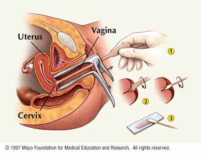
  • ก่อนไปตรวจก็ควรทำความสะอาดภายนอกโดยใช้สบู่ธรรมดา ไม่ต้องใส่น้ำหอมหรือกลิ่น
  • ควรสวมกระโปงหรือกางเกงหลวมๆที่สามารถถอดออกง่าย
  • ไม่ควรจะไปเล่นกีฬาหรือไป shopping ก่อนการตรวจ
  • ควรจะงดการมีเพศสัมพันธุ์ก่อนการตรวจ
  • ไม่ควรสวนหรือล้างภายในช่องคลอด
  • ไม่ควรจะเหน็บยา
  • ไม่ควรไปตรวจช่วงมีประจำเดือน

วิธีการตรวจ

เมื่อท่านได้แจ้งกับเจ้าหน้าที่ว่าต้องการจะตรวจภายใน เจ้าหน้าที่ก็จะนำท่านไปพบแพทย์หลังจากซักประวัติ และตรวจร่างกายแล้วก็จะเข้าห้องตรวจภายในซึ่งเป็นห้องที่มิดชิดพอสมควร เจ้าหน้าที่จะแนะนำให้ท่านเปลี่ยนกางเกงหรือกระโปงเป็นผ้าที่มีลักษณะเหมือนผ้าถุง เมื่อเปลี่ยนเสื้อผ้าเสร็จแล้ว เจ้าหน้าที่จะนำท่านไปยังเตียงตรวจซึ่งไม่เหมือนกับเตียงตรจที่ท่านเคยเห็น เมื่อนอนเตียงเรียบร้อยแล้วเจ้าหน้าที่จะให้ท่านวางเท้าไว้บนขาหยั่งซึ่งจะทำให้ท่านต้องแยกขาออก เจ้าหน้าที่จะเปิดผ้าถุง และนำผ้ามาคลุมและเปิดช่องไว้เพียงพอในการตรวจ หลังจากนั้นจะเรียกแพทย์มาตรวจ แพทย์จะทำความสะอาดบริเวณดังกล่าวด้วยน้ำยาฆ่าเชื้อ แล้วจึงใส่ speculum เพื่อขยายช่องคลอด การเลือด speculum แพทย์จะอาศัยประวัติการมีเพศสัมพันธ์และประวัติการคลอดบุตร การใส่ speculum อาจจะสร้างความอึกอัดเล็กน้อย หลังจากนั้นแพทย์จะใช้ไม้ไปขูดเนื้อเยื่อที่ปากมดลูก และนำเซลล์นั้นไปส่งตรวจหามะเร็ง

การแปลผล

หลังจากที่แพทย์ส่งเซลล์ไปตรวจต้องใช้เวลารอสัก 2-3 วันผลรายงานที่ออกมามีดังนี้

  • ปกติหรือ Normal หมายถึงตรวจไม่พบเซลล์ที่ผิดปกติ ดังนั้นไม่ต้องทำอะไรต่อ
  • Atypical squamous cells of undetermined significance มีความผิดปกติของเซลล์แต่ไม่สามารถบอกได้อย่างแน่ชัดว่าเป็นมะเร็งหรือไม่ อาจจะเกิดจากการติดเชื้อหรือการอักเสบของปาดมดลูก แพทย์จะแนะนำให้มาตรวจซ้ำ
  • Squamous intraepithelial lesion ลักษณะที่ตรวจพบจะมีโอกาศเป็นมะเร็งได้สูง แพทย์จะต้องนัดตรวจเพิ่มเติม
  • Atypical glandular cellsหมายถึงมีเซลล์ที่ผลิตมูกมาก หากได้รับรายงานนี้แพทย์ต้องตรวจต่อ
  • SILLow-grade Squamous Intraepithelial Lesion.หมายถึงมีการเปลี่ยนแปลงขนาดและรูปร่างของเซลล์ ภาวะนี้เกิดจากการติดเชื้อ HPV ผู้ป่วยส่วนใหญ่จะหายเป็นปกติ
  • HSILHigh-grade Squamous Intraepithelial Lesion. มีการเปลี่ยนแปลงขนาดและรูปร่างของเซลล์ที่ชัดเจน หากไม่รักษาจะกลายเป็นมะเร็ง
  • Squamous cancer or adenocarcinoma cells ผลออกมาแบบนี้ก็ต้องตรวจต่อว่าเป็นมะเร็งมากแค่ไหน เพื่อวางแผนการรักษา

The Papanicolaou (Pap) test

The Pap test (Pap smear) is the main screening test for cervical cancer and pre-cancerous changes.

Although the Pap test has been more successful than any other screening test in preventing a cancer, it is not perfect. One of the limitations of the Pap test is that it needs to be examined by humans, so an accurate analysis of the hundreds of thousands of cells in each sample is not always possible. Engineers, scientists, and doctors are working together to improve this test. Because some abnormalities may be missed (even when samples are looked at in the best laboratories), it is not a good idea to have this test less often than American Cancer Society guidelines recommend.

Pelvic exam versus Pap test

Many people confuse pelvic exams with Pap tests. The pelvic exam is part of a woman's routine health care. During a pelvic exam, the doctor looks at and feels the reproductive organs, including the uterus and the ovaries and do tests for sexually transmitted disease. Pap tests are often done during pelvic exams, but you can have a pelvic exam without having a Pap test. A pelvic exam without a Pap test will not help find cervical cancer at an early stage or find abnormal cells of the cervix.

The Pap test is often done at the start of the pelvic exam, after the speculum is placed. To do a Pap test, the doctor must remove cells from the cervix by gently scraping or brushing with a special instrument. Pelvic exams may help find other types of cancers and reproductive problems, but only Pap tests give information on early cervical cancer or precancers.

How the Pap test is done

Cytology is the branch of science that deals with the structure and function of cells. It also refers to tests to diagnose cancer by looking at cells under the microscope. The Pap test (or Pap smear) is a procedure used to collect cells from the cervix for cervical cytology testing.

The health care professional first places a speculum inside the vagina. The speculum is a metal or plastic instrument that keeps the vagina open so that the cervix can be seen clearly. Next, using a small spatula, a sample of cells and mucus is lightly scraped from the exocervix (the surface of the cervix that is closest to the vagina). A small brush or a cotton-tipped swab is then used to take a sample from the endocervix (the inside part of the cervix that is closest to the body of the uterus). There are 2 main ways to prepare the cell samples so that they can be examined under a microscope in the laboratory.

  1. Conventional cytology: The first way is to smear the sample directly onto a glass microscope slide, which is then sent to the laboratory. For about 50 years, all cervical cytology samples were handled this way. This method works quite well and is relatively inexpensive, but it does have some drawbacks. One problem with this method is that the cells smeared onto the slide are sometimes piled up on each other, making it hard to see the cells at the bottom of the pile. Also, white blood cells (pus), increased mucus, yeast cells, or bacteria from infection or inflammation can hide the cervical cells. Another problem with direct smears is that if the slides are not treated (with a preservative) right away, the cells can dry out. This can make it difficult to tell if there is something wrong with the cells. If the cervical cells cannot be seen well (because of any of these problems), the Pap smear may need to be done again.

  2. Liquid based cytology: Another method is to put the sample of cells from the cervix into a special preservative liquid (instead of putting them on a slide directly). Technicians use special lab instruments that spread the cells in the liquid onto glass slides to look at under the microscope. This method is called liquid-based cytology, or a liquid-based Pap test. The liquid helps remove some of the mucus, bacteria, yeast, and pus cells in a sample. It also allows the cervical cells to be spread more evenly on the slide and keeps them from drying out and distorting. Cells kept in the liquid can also be tested for HPV. Using liquid-based testing reduces the chance that the Pap test will need to be repeated, but it does not seem to find more pre-cancers than a regular Pap test. This method, also known by brand names ThinPrep® or AutoCyte®, is more expensive than a usual Pap test.

Another way to improve the Pap test is by using computerized instruments that can spot abnormal cells in Pap tests. The AutoPap® instrument has been approved by the FDA to read Pap tests first (instead of them being examined by a technologist). It is also approved by the FDA for rechecking Pap test results that were read as normal by technologists. Any smear identified as abnormal by the AutoPap® would then be reviewed by a doctor or a technologist.

Computerized instruments can find abnormal cells that technologists sometimes miss. Most of the abnormal cells found in this way are in rather early stages, such as atypical squamous cells (ASCs), but sometimes high-grade abnormalities missed by human testing can be found. Scientists do not know yet whether the instrument can find enough high-grade abnormalities missed by human testing to have a real impact on preventing invasive cervical cancers. Automated testing also increases the cost of the cervical cytology testing.

For now, the best way to detect cervical cancer early is to make certain that all women are tested according to American Cancer Society guidelines. Unfortunately, many of the women most at risk for cervical cancer are not being tested often enough or at all.

Making your Pap tests more accurate

You can do several things to make your Pap test as accurate as possible:

  • Try not to schedule an appointment for a time during your menstrual period. The best time is at least 5 days after your menstrual period stops.
  • Don't use tampons, birth-control foams, jellies or other vaginal creams for 2 to 3 days before the test.
  • Don't douche for 2 to 3 days before the test.
  • Don't have sexual intercourse for 2 days before the test.

Is there a "home" Pap test?

Doctors have been trying to find ways to get more women involved in cervical cancer screening. Some have proposed methods that would allow women to obtain cervical cell samples at home. For this test, a woman would collect cervical cells herself by inserting a small plastic applicator into the vagina and moving it around on the cervix. This would be placed in a special container to preserve the cells. This type of approach has been used to check for sexually transmitted diseases in poorer countries, and it has also been useful to check for HPV infections. So far, however, no "home-based" Pap test has been approved for use in the U.S. Currently, the American Cancer Society does not recommend any "at home" Pap test.

How Pap test results are reported

The most widely used system for describing Pap test results is the Bethesda System (TBS). This system has been revised twice since it was developed in 1988 -- first in 1991 and, most recently, in 2001. The information that follows is based on the 2001 version. There are 3 main categories, some of which have sub-categories:

  • negative for intraepithelial lesion or malignancy,
  • epithelial cell abnormalities, and
  • other malignant neoplasms.

Negative for intraepithelial lesion or malignancy: This first category means that no signs of cancer, precancerous changes, or other significant abnormalities were found. Some specimens in this category appear entirely normal. Others may have findings that are unrelated to cervical cancer, such as signs of infection (with yeast, herpes, or trichomonas, for example). Some cases may also show "reactive cellular changes", which the way cervical cells respond to infection or other irritation.

Epithelial cell abnormalities: The second category, epithelial cell abnormalities, means that the cells of the lining layer of the cervix show changes that might be cancer or a pre-cancerous condition. This category is divided into several groups for squamous cells and glandular cells. The epithelial cell abnormalities for squamous cells are:

Atypical squamous cells (ASCs): This term is used when there are cells that look abnormal, but it is not possible to tell (by looking at the cells under a microscope) if this caused by infection, irritation, or precancer. This group is further divided into 2 groups, atypical squamous cells of uncertain significance (ASC-US) and atypical squamous cells where high-grade squamous intraepithelial lesion (SIL) can’t be excluded (ASC-H). Most of the time, cells labeled ASC-US are not pre-cancer.. Some doctors will recommend repeating the Pap test after several months. Some doctors use the HPV DNA test to help them decide on the best treatment plan. If a woman with ASC-US is infected with a high-risk type of HPV, doctors are more inclined to do a colposcopy. The term ASC-H is used if a SIL is suspected. Colposcopy is usually done in cases of ASC-H.

Squamous intraepithelial lesions (SILs): These abnormalities are subdivided into low-grade SIL and high-grade SIL. All patients with this Pap test result should have colposcopy. High-grade SILs are less likely than low-grade SILs to go away without treatment. They are also more likely to eventually develop into cancer if they are not treated. Treatment can cure all SILs and prevent true cancer from developing. A Pap test cannot tell for certain whether a woman has a high- or low-grade SIL. It merely flags the result as fitting into one of these abnormal categories. All patients with a Pap test result of SIL should have colposcopy. The need for treatment is based on the results of the colposcopy. Since most SILs are positive for HPV, HPV testing is not very helpful in deciding what to do for an SIL.

Squamous cell carcinoma: This result means that the woman is likely to have an invasive squamous cell cancer. Further testing will be done to be sure of the diagnosis beforetreatment can be planned.

The Bethesda System also describes epithelial cell abnormalities for glandular cells.

Adenocarcinoma: Cancers of the glandular cells are called adenocarcinomas. In some cases, the pathologist examining the cells can suggest whether the adenocarcinoma started in the endocervix, in the uterus (endometrium), or elsewhere in the body.

Atypical glandular cells: When the glandular cells do not look normal, but have features that do not permit a clear decision as to whether they are cancerous, the term used is atypical glandular cells (AGCs). The patient will usually have more testing if her cervical cytology result shows atypical glandular cells.

Other malignant neoplasms: This category refers to forms of cancer that rarely affect the cervix, such as malignant melanoma, sarcomas, and lymphoma.

Other methods of describing Pap test results have also been used in the past.

The HPV DNA Test

As mentioned earlier, the most important risk factor for developing cervical cancer is infection with HPV. Doctors can now test for the types of HPV that are most likely to cause cervical cancer ("high-risk" or "carcinogenic" types) by looking for pieces of their DNA in cervical cells. The test is done in a similar way to the Pap test in terms of how the sample is collected, and in some cases can even be done on the same sample.

The HPV DNA test can be used in 2 situations:

  • The FDA recently approved the HPV gene test to be used combination with the Pap test to screen for cervical cancer in women over 30 years old (see American Cancer Society screening guidelines above). It does not replace the Pap test.. Women in their 20s who are sexually active are much more likely (than older women) to have an HPV infection that will go away on its own. For these younger women, results of this test are not as significant and may be more confusing. . For this reason, the HPV DNA test is not recommended as a screening test in women under 30. For more information, see the American Cancer Society documents, What Every Woman Should Know About Cervical Cancer and the Human Papilloma Virus and Thinking About Testing for HPV?
  • The HPV DNA test is also used in women of any age who have slightly abnormal Pap test results to find out if they might need more testing or treatment. (see next section).

Other tests for women with abnormal cervical cytology results

The Pap test is a screening test, not a diagnostic test - it cannot tell for certain that cancer is present. An abnormal Pap test result means that you will likely need to have other tests to find out if a cancer or a pre-cancer is actually present. The tests that are used include colposcopy (with biopsy) and endocervical scraping. These tests are commonly used when the results of a Pap test are reported as SIL, atypical glandular cells, or cancer. If a biopsy shows a pre-cancer, doctors will take steps to keep an actual cancer from developing.

Doctors are less certain about what to do when the Pap test result shows atypical squamous cells. In deciding what to do, some doctors take into account your previous Pap test results, whether you have any cervical cancer risk factors, whether you have remembered to have Pap tests done in the past, and whether the test result is ASC-H or ASC-US. For ASC-H, many doctors will recommend colposcopy and biopsy. For ASC-US some doctors will recommend colposcopy and biopsy if a high-risk HPV DNA is detected, but others recommend repeating the Pap test after several months.

Colposcopy

If you have certain symptoms that suggest cancer or if your Pap test shows abnormal cells, you will need to have a test called colposcopy. In this procedure you will lie on the exam table as you do with a pelvic exam. A speculum will be placed in the vagina to help the doctor see the cervix. The doctor will use the colposcope to examine the cervix. The colposcope is an instrument with magnifying lenses (like binoculars), that lets the doctor see the surface of the cervix closely and clearly. The doctor may "treat" your cervix with a weak solution of acetic acid (similar to vinegar) to make any abnormal areas easier to see.

Colposcopy is not painful, has no side effects, and can be done safely even if you are pregnant. If an abnormal area is seen on the cervix, a biopsy will be done. For a biopsy, small piece of tissue is removed from the area that looks abnormal. The sample is sent to a pathologist to look at under a microscope. A biopsy is the only way to tell for certain whether an abnormal area is a pre-cancer, a true cancer, or neither.

Cervical biopsies

Several types of biopsies are used to diagnose cervical precancers and cancers. If the biopsy can completely remove all of the abnormal tissue, it may be the only treatment needed. In some situations, additional treatment of precancers or cancers is needed.

Colposcopic biopsy: For this type of biopsy, a doctor or other health care professional first examines the cervix with a colposcope to find the abnormal areas. Using a biopsy forceps, the doctor will remove a small (about 1/8-inch) section of the abnormal area on the surface of the cervix. The biopsy procedure may cause mild cramping or brief pain, and you may have light bleeding afterward. A local anesthetic is sometimes used to numb the cervix before the biopsy.

Endocervical curettage (endocervical scraping): The colposcope does not help the doctor see into the endocervix. The endocervix will have to be scraped to see if it is affected by pre-cancer or cancer. This procedure is usually done at the same time as the colposcopic biopsy. A local anesthetic may be used to numb the cervix. Then, a narrow instrument (called a curette) is inserted into the endocervical canal (the passage between the outer part of the cervix and the inner part of the uterus). Some of the tissue that is lining the endocervical canal is removed by scraping with the curette. This tissue sample is sent to the lab for examination. After this procedure, patients may feel a cramping pain, and they may also have some light bleeding.

Cone biopsy: In this procedure, also known as conization, the doctor removes a cone-shaped piece of tissue from the cervix. The base of the cone is formed by the exocervix (outer part of the cervix), and the point or apex of the cone is from the endocervical canal. The transformation zone (the border between the exocervix and endocervix) is contained within the cone. This is the area of the cervix where pre-cancers and cancers are most likely to develop. The cone biopsy can be used as a treatment to completely remove many pre-cancers and some very early cancers. Having a cone biopsy will not keep most women from getting pregnant, but if the biopsy removes large amount of tissue these women may have a higher risk of giving birth prematurely.

There are 2 methods commonly used for cone biopsies: the loop electrosurgical excision procedure (LEEP; also called large loop excision of the transformation zone [LLETZ]) and the cold knife cone biopsy.

  • LEEP (LLETZ): With this method, the tissue is removed with a thin wire loop that is heated by electrical current and acts as a scalpel. For this procedure, a local anesthetic is used, and it can be done in your doctor's office. It can take as little as 10 minutes. You may have mild cramping during and after the procedure, and mild to moderate bleeding may persist for several weeks.
  • Cold knife cone biopsy: This method uses a surgical scalpel or a laser instead of a heated wire to remove tissue. It requires general anesthesia (you are asleep during the operation) and is done in a hospital, but no overnight stay is needed. After the procedure, cramping and some bleeding may persist for a few weeks.

How biopsy results are reported: The terms for reporting biopsy results are slightly different from the Bethesda System for reporting Pap test results. Pre-cancerous changes are called "cervical intraepithelial neoplasia" (CIN) or, rarely, "dysplasia," instead of "squamous intraepithelial lesion" (SIL). The terms for reporting cancers ("squamous cell carcinoma" and "adenocarcinoma") are the same.

How women with abnormal Pap test results are treated to prevent cervical cancers from developing

If an abnormal area is seen during the colposcopy, your doctor will be able to remove it with LEEP (LLETZ procedure). Other options include a cold knife cone biopsy and destroying the abnormal cells with cryosurgery or laser surgery.

During cryosurgery, the doctor uses a metal probe cooled with liquid nitrogen to kill the abnormal cells by freezing them.

In laser surgery, the doctor uses a focused beam of high-energy light to vaporize (burn off) the abnormal tissue. This is done through the vagina, with local anesthesia.

Both cryosurgery and laser surgery can be done in a doctor's office or clinic. After treatment, you may have alot of watery brown discharge for a few weeks.

These treatments are almost always effective in destroying pre-cancers and preventing them from developing into true cancers. You will need follow-up exams to make sure that the abnormality does not come back. If it does, the treatments can be repeated

เมื่อได้รับรายว่าว่าผลการตรวจ Pap แพทย์จะนัดมาตัดชิ้นเนื้อที่ปากมดลูกเพื่อการวินิจฉัยที่แน่นอน